Interpreting “Biology Corner” Resources
Animal cell coloring biology corner answer key – Biology Corner is a valuable online resource for educators and students alike, offering a wealth of information on various biological topics, including animal cells. Understanding how to effectively utilize these resources, particularly in conjunction with hands-on activities like coloring, is key to maximizing their educational impact.The typical information presented in Biology Corner’s animal cell resources usually includes detailed diagrams showcasing the cell’s organelles, descriptions of each organelle’s structure and function, and often, interactive quizzes or worksheets to assess comprehension.
These resources frequently incorporate engaging visuals and clear, concise language, making them accessible to a wide range of learners.
Using Biology Corner Resources with Coloring Activities, Animal cell coloring biology corner answer key
Biology Corner’s resources can be seamlessly integrated with coloring activities to enhance learning and retention. For example, students can first study the diagrams and descriptions of animal cell organelles on Biology Corner. Then, they can use a blank animal cell diagram or a worksheet provided by Biology Corner (or create their own) to color-code each organelle based on its function or characteristics.
This active learning approach strengthens memory and understanding by connecting visual representation with textual information. The coloring activity acts as a reinforcement exercise, solidifying the knowledge gained through reading and studying the online material.
Comparison of Approaches to Teaching Animal Cell Structure
Different educational materials employ diverse approaches to teaching animal cell structure. Some materials focus heavily on rote memorization of organelles and their functions, often using lists and simple diagrams. Others utilize more interactive methods, such as animations, virtual labs, or 3D models, allowing students to explore the cell structure in a more dynamic way. Biology Corner, in this context, offers a middle ground, providing both textual information and visual aids but also leaving room for creative integration with other teaching methods, like the aforementioned coloring activity.
A purely text-based approach might prove less engaging for visual learners, while a solely animation-based approach may lack the depth of detail provided by text-based resources. The effectiveness of each approach often depends on the learning style of the student and the specific learning objectives. A balanced approach, incorporating multiple methods, usually yields the best results.
Creating a Detailed Animal Cell Illustration: Animal Cell Coloring Biology Corner Answer Key

Illustrating an animal cell requires careful consideration of its three-dimensional structure and the arrangement of its various organelles. This description will guide the creation of a detailed and accurate representation, suitable for educational purposes. We will focus on accurately depicting the organelles’ relative sizes and positions within the cell’s cytoplasm.
Finding the answer key for your animal cell coloring worksheet from Biology Corner can be tricky, but expanding your search to include broader resources can be helpful. For a more comprehensive approach to cell structure, you might find a useful resource in this animal and plant cell coloring pdf , which offers a visual comparison. This broader perspective can then aid in understanding the specifics of the animal cell coloring assignment and its associated answer key.
The animal cell, unlike plant cells, lacks a rigid cell wall and a large central vacuole. This allows for a more flexible and irregular shape. The cell’s overall appearance is that of a roughly spherical or oval structure, though its precise shape can vary depending on its function and surroundings.
Organelle Representation and Arrangement
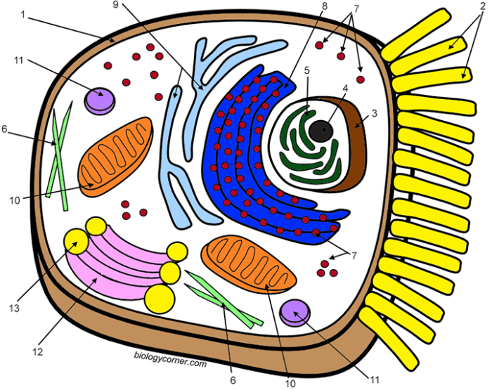
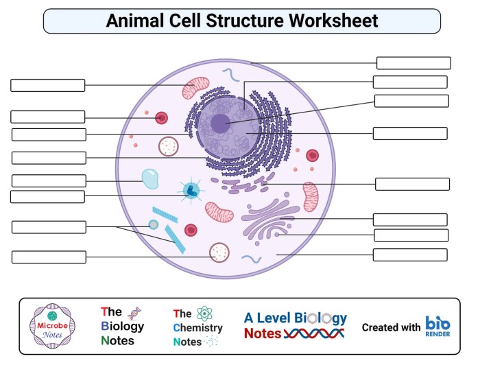
To accurately represent an animal cell, it’s crucial to understand the spatial relationships between organelles. The nucleus, typically the largest organelle, should be centrally located or slightly off-center. The endoplasmic reticulum (ER) extends throughout the cytoplasm as a network of interconnected membranes, both smooth and rough (the rough ER having ribosomes attached). The Golgi apparatus, often depicted as a stack of flattened sacs (cisternae), is usually positioned near the nucleus.
Mitochondria, the powerhouses of the cell, are scattered throughout the cytoplasm, often depicted as bean-shaped structures with internal cristae. Lysosomes, small spherical vesicles containing digestive enzymes, are distributed more randomly.
Depicting Three-Dimensionality in Two Dimensions
Representing the three-dimensional nature of an animal cell on a two-dimensional surface requires careful use of shading, perspective, and overlapping organelles. Organelles closer to the “foreground” of the drawing should be more sharply defined and detailed, while those further “back” can be slightly less defined and may overlap partially with other organelles. Shading can be used to create a sense of depth and volume, suggesting the curvature of the cell membrane and the three-dimensional structure of organelles like mitochondria.
Overlapping organelles can effectively communicate their relative positions within the cell. For example, the endoplasmic reticulum should be depicted weaving between and around other organelles, highlighting its extensive network. The Golgi apparatus should be drawn with a sense of depth, showcasing the stacked nature of the cisternae.
Specific Organelle Details
The following details will aid in the accurate representation of each organelle:
- Nucleus: A large, roughly spherical structure containing the genetic material (DNA). Depict a nuclear envelope (double membrane) with nuclear pores. The nucleolus, a smaller, denser region within the nucleus, should also be shown.
- Ribosomes: Small, granular structures either free-floating in the cytoplasm or attached to the rough endoplasmic reticulum. Represent them as small dots or slightly elongated shapes.
- Endoplasmic Reticulum (ER): A network of interconnected membranes extending throughout the cytoplasm. The rough ER should be depicted with ribosomes attached, giving it a rough appearance. The smooth ER should be shown as a network of smooth membranes.
- Golgi Apparatus: A stack of flattened, membrane-bound sacs (cisternae). Illustrate the cisternae as slightly curved and overlapping, showing the three-dimensional structure.
- Mitochondria: Bean-shaped organelles with a double membrane. The inner membrane should be folded into cristae, which should be depicted as folds or ridges within the mitochondrion.
- Lysosomes: Small, spherical vesicles containing digestive enzymes. Depict them as small, uniformly colored circles.
- Centrioles: A pair of cylindrical structures involved in cell division. Show these as short, cylindrical structures usually located near the nucleus.
- Cell Membrane: The outer boundary of the cell. Illustrate it as a thin, continuous line, subtly suggesting a curved surface.
Analyzing Common Errors in Animal Cell Diagrams

Students often encounter difficulties when illustrating animal cells, leading to inaccuracies in their diagrams. These inaccuracies stem from a lack of understanding of the cell’s components, their relative sizes, and their spatial arrangements. Addressing these misconceptions is crucial for building a solid foundation in cell biology. Interactive learning activities can effectively correct these misunderstandings and promote accurate representations.Common Misconceptions in Animal Cell Diagrams and Their Correction
Incorrect Representation of Organelles
Students frequently misrepresent the size, shape, and location of organelles within the animal cell. For instance, the nucleus is often drawn too small relative to the cytoplasm, or the Golgi apparatus is depicted as a simple, singular structure rather than a complex network of flattened sacs. Mitochondria might be shown as uniform circles instead of their characteristic bean shape.
To correct this, interactive activities like building a 3D model of an animal cell using readily available materials (e.g., modeling clay, beads) can help students visualize the relative sizes and spatial relationships of different organelles. Additionally, comparing student drawings to accurate microscopic images will highlight discrepancies and reinforce correct representations.
An inaccurate representation might show a tiny nucleus squeezed into a corner of a large, empty cell, while an accurate representation would depict a relatively large, centrally located nucleus occupying a significant portion of the cell’s volume.
An inaccurate depiction might show the mitochondria as small, uniform spheres, whereas an accurate one would illustrate them as elongated, bean-shaped structures with internal cristae.
Omission or Incorrect Placement of Key Organelles
Another common error involves the omission of essential organelles or their incorrect placement within the cell. Lysosomes, ribosomes, and the endoplasmic reticulum are frequently forgotten or incorrectly positioned. To address this, interactive games or quizzes that require students to identify and locate organelles within a virtual animal cell can be highly effective. These activities can be further enhanced by incorporating drag-and-drop features or labeling exercises.
This gamified approach makes learning engaging and reinforces accurate knowledge of organelle location and function.
Incorrect Labeling of Cell Structures
Incorrect or missing labels are frequent issues in student diagrams. Students might mislabel organelles or incorrectly associate functions with structures. To improve labeling accuracy, students can participate in peer review sessions where they critique each other’s diagrams and provide constructive feedback on labeling accuracy. This fosters collaboration and promotes a deeper understanding of the functions of each organelle.
Furthermore, providing a detailed checklist of organelles and their functions can serve as a helpful guide during the drawing and labeling process.
Inconsistent Scale and Proportion
Students often fail to maintain consistent scale and proportion when drawing animal cells. Organelles might be disproportionately large or small compared to each other and the cell itself. To remedy this, using pre-made templates with scaled grids can assist students in accurately representing the relative sizes of different organelles. The use of microscopic images as a reference point can further enhance the accuracy of scale and proportion in their drawings.
Query Resolution
What are some alternative methods for teaching animal cell structure besides coloring?
Model building, virtual reality simulations, and interactive whiteboard activities are effective alternatives.
Where can I find additional resources similar to “Biology Corner”?
Websites like Khan Academy, CK-12, and educational YouTube channels offer comparable resources.
How can I assess student understanding beyond just coloring activities?
Use quizzes, tests, labeled diagrams, and short essays to assess comprehension.
Are there any specific software or apps that can aid in creating interactive animal cell diagrams?
Several software options and apps exist, including BioRender, Adobe Illustrator, and various educational apps.
