Coloring Sheet Design and Functionality: Animal Cell Coloring Sheets Answers

Animal cell coloring sheets answers – This section details the design and functionality of a simplified animal cell coloring sheet, aiming to make learning about cell organelles engaging and effective. The design prioritizes visual clarity and accurate representation of key cellular components. The color choices are intended to be both visually appealing and mnemonically helpful, associating specific colors with specific organelles. Suggestions for using the coloring sheet as a learning tool are also included.
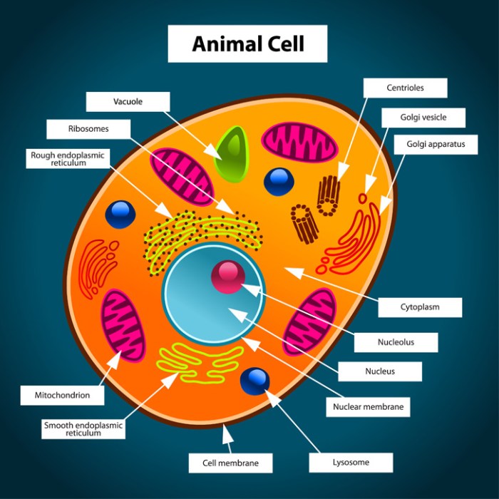
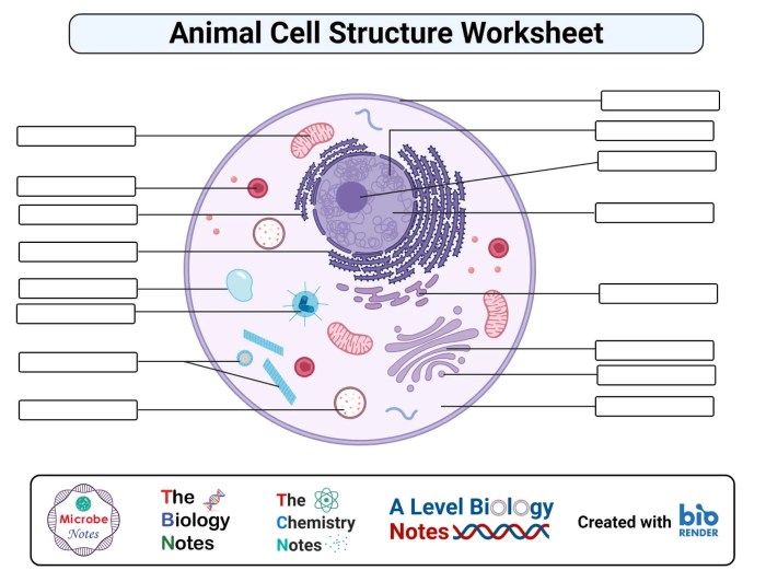
The coloring sheet depicts a simplified animal cell, focusing on the major organelles. The cell’s structure is presented in a manner easily understandable for younger learners while remaining scientifically accurate. The organelles are clearly labeled, making it easy to identify and color them correctly. This simplified representation avoids overwhelming detail, focusing on core components and their functions.
Organelle Representation and Color Choices
Selecting appropriate colors for each organelle aids in memorization and visual differentiation. The following list details the organelles and the suggested colors, along with reasoning for the choice.
Finding the answers for your animal cell coloring sheets can be tricky, but a great way to learn is by using a printable coloring page first. You can find a helpful resource for this at animal cell coloring page printable , which will allow you to visually identify the different organelles before tackling the answers. Once you’ve colored and labeled the page, checking your animal cell coloring sheets answers will be much easier.
- Cell Membrane: Light Blue. This represents the fluid and flexible nature of the membrane, suggesting movement and interaction.
- Cytoplasm: Light Yellow. This pale color provides a neutral background, allowing the organelles to stand out clearly.
- Nucleus: Dark Purple. The dark color highlights the nucleus’s importance as the cell’s control center.
- Nucleolus: Darker shade of Purple. The slightly darker shade distinguishes the nucleolus within the nucleus.
- Ribosomes: Dark Green. The dark color represents the ribosomes’ vital role in protein synthesis, a key cellular function.
- Endoplasmic Reticulum (ER): Light Green (Rough ER) and Light Pink (Smooth ER). The difference in color helps distinguish the rough and smooth ER, highlighting their distinct functions.
- Golgi Apparatus: Orange. The vibrant color makes the Golgi apparatus easily identifiable, emphasizing its role in packaging and processing proteins.
- Mitochondria: Red. Red signifies energy, reflecting the mitochondria’s function as the powerhouse of the cell.
- Lysosomes: Dark Brown. This color symbolizes the lysosomes’ role in breaking down waste materials.
- Vacuoles: Light Pink (multiple small vacuoles). The color choice distinguishes them from the smooth ER and highlights their storage function.
Utilizing the Coloring Sheet as a Learning Tool
The coloring sheet can be effectively integrated into various learning activities. The act of coloring itself encourages engagement and retention of information.
- Labeling Activity: Students can label the organelles after coloring, reinforcing their names and locations.
- Function Matching: Students can match the colored organelles to their functions written on separate cards.
- Comparative Study: The coloring sheet can be used to compare and contrast animal cells with plant cells (if a similar plant cell coloring sheet is available).
- Presentation Tool: Colored sheets can be used as visual aids during presentations or classroom discussions.
Organizing Coloring Sheet Elements for Optimal Clarity
The layout of the coloring sheet is crucial for its effectiveness. A clear and organized presentation enhances understanding.
The animal cell should be centrally located, with organelles proportionally sized and clearly separated. Labels for each organelle should be placed directly next to or slightly above the corresponding organelle, using a clear and legible font. A title, “Animal Cell,” should be prominently displayed at the top. The overall design should be visually appealing and avoid clutter. Using a light background color enhances visibility and reduces eye strain.
Illustrative Examples and Descriptions

This section provides detailed descriptions of key organelles within an animal cell, focusing on their visual representation in a coloring sheet and their functional roles. Clear and concise descriptions will aid in understanding the cell’s intricate workings.
Mitochondrion
The mitochondrion, often depicted as a bean-shaped structure with a folded inner membrane, is the powerhouse of the cell. In the coloring sheet, it could be represented as a bean or oval shape with internal lines illustrating the cristae, the folds of the inner membrane. These cristae significantly increase the surface area for cellular respiration. The mitochondrion’s primary function is to generate adenosine triphosphate (ATP), the cell’s main energy currency, through the process of cellular respiration.
This involves breaking down glucose and other nutrients in the presence of oxygen to produce ATP, which fuels various cellular processes. The coloring sheet illustration should clearly show the double membrane structure—an outer smooth membrane and an inner folded membrane—to accurately reflect its complex architecture.
Nucleus, Animal cell coloring sheets answers
The nucleus, the cell’s control center, is typically depicted as a large, round structure. The coloring sheet representation should include a clearly defined nuclear membrane (or nuclear envelope), a double membrane that separates the nucleus from the cytoplasm. Within the nucleus, a darker area should represent the nucleolus, the site of ribosome production. The caption could read: “The Nucleus: The cell’s control center, containing the genetic material (DNA) organized into chromosomes.
The nucleolus, a dense region within the nucleus, is responsible for ribosome synthesis.” The dispersed chromatin, representing the less condensed form of DNA, should also be visible within the nucleus.
Golgi Apparatus
The Golgi apparatus, often visualized as a stack of flattened sacs or cisternae, is crucial for protein modification, sorting, and packaging. In the coloring sheet, it can be represented as a series of interconnected, flattened membrane-bound sacs. Small vesicles, representing the transport containers for proteins, should be shown budding off from the Golgi’s edges. Its function is to receive proteins synthesized by the endoplasmic reticulum, modify them (e.g., glycosylation), sort them, and package them into vesicles for transport to their final destinations within or outside the cell.
The visual representation should clearly depict the stacked structure and the budding vesicles to illustrate this dynamic process.
Endoplasmic Reticulum
The endoplasmic reticulum (ER) is a network of interconnected membranes extending throughout the cytoplasm. In the coloring sheet, it can be illustrated as a series of interconnected, branching tubules and flattened sacs. The rough endoplasmic reticulum (RER), studded with ribosomes (small dots), should be distinguished from the smooth endoplasmic reticulum (SER), which lacks ribosomes. The RER is primarily involved in protein synthesis, while the SER plays a significant role in lipid metabolism and detoxification.
The coloring sheet should visually differentiate the RER and SER to highlight their distinct roles. The overall network structure should convey its extensive presence within the cell and its role in transporting proteins and lipids.
Helpful Answers
What are the best materials to use for coloring the animal cell?
Crayons, colored pencils, markers, or even paint can be used. The choice depends on the age and preference of the user.
Can these coloring sheets be used for homeschooling?
Absolutely! They are an excellent resource for homeschooling, providing a hands-on and engaging way to teach cell biology.
How can I assess student understanding after using the coloring sheets?
Use accompanying worksheets, quizzes, or oral questioning to assess comprehension. Observe the accuracy of organelle labeling and coloring on the sheet itself.
Are there any online resources available for printable animal cell coloring sheets?
Yes, many websites offer free printable animal cell coloring sheets. A simple online search should yield numerous results.
